
Tumours of the Larynx
BENIGN TUMOURS
Benign tumours of the larynx are rare and cause persistent hoarseness.The
commonest tumours encountered are:
1 papilloma—solitary or multiple;
2 haemangioma—almost exclusively in infants;
3 fibroma.
Papillomata and haemangiomata are considered in more detail in Chapter
37.
MALIGNANT TUMOURS
PATHOLOGY
Malignant tumours of the larynx are virtually always squamous cell carcinoma.
Adenoid cystic carcinoma and sarcoma may occur on rare
occasions.
AETIOLOGY
Malignant tumours are commoner in males by a ratio of 10:1, occurring
almost exclusively in smokers. The peak age incidence is 55–65 years, but
it can occur in young adults.
Glottic carcinoma (60% of cases)
The prime symptom of glottic carcinoma (Fig. 35.1) is hoarseness,
which may persist as the only symptom for many months. Only when
spread from the cord has occurred will earache, dysphagia and dyspnoea
supervene.
Supraglottic carcinoma (30% of cases)
Supraglottic carcinoma, as well as producing a change in the voice, may
metastasize early to the cervical nodes.
Subglottic carcinoma
Subglottic carcinoma produces less hoarseness but increasing airway obstruction.
It must not be mistaken for asthma or chronic bronchitis.
SPREAD OF LARYNGEAL CARCINOMA
Spread is local initially and proceeds:
1 along the cord to the anterior commissure and onto the opposite cord;
2 upwards onto the ventricular band and epiglottis;
3 downwards to the subglottis;
4 deeply into the laryngeal muscles, causing cord fixation.
Lymphatic spread from glottic lesions is late, but occurs readily from supraglottic
and subglottic sites to the deep cervical nodes.
Pulmonary metastases occur occasionally but other distant metastases
are rare.
DIAGNOSIS
Every case of hoarseness should be examined by indirect laryngoscopy;
malignant growths are usually seen easily. Diagnosis is confirmed by microlaryngoscopy
and biopsy.
The chest must be X-rayed as bronchial carcinoma also may be present.
CT scanning of the larynx is often helpful in defining the extent of spread,
and is usually performed prior to deciding on treatment.
TREATMENT
1 Radiotherapy by external radiation is usually employed. In a small tumour
limited to one cord (the stage at which it should be diagnosed), the 5-
year survival rate is 80–90% and the patient retains a normal larynx.
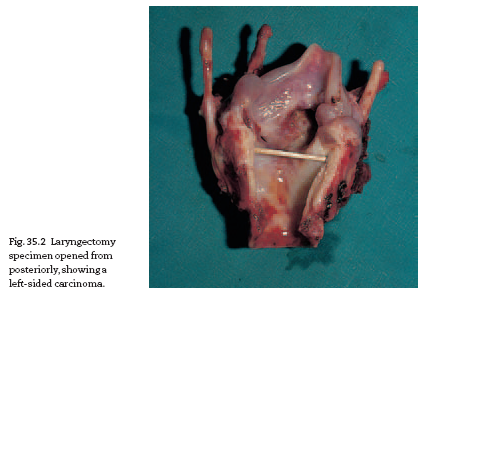
2 In very extensive disease or if there is recurrence following radiotherapy,
total laryngectomy is necessary (Fig. 35.2).The patient obviously then
has a permanent tracheostomy and will need to develop oesophageal
speech. Good oesophageal speech is attained by about 30% of patients; a
further 30% develop reasonable voice but the remainder never manage
more than a mouthed whisper.
Many patients are now provided with a tracheopharyngeal valve. A
fistula is formed between the trachea and pharynx and a prosthetic valve
fitted to the fistula. Occlusion by the finger of the tracheostomy allows
air to flow into the hypopharynx, while vibration of the soft tissue produces
phonation. This then allows fluent lung-powered voice for the
laryngectomee.
Rehabilitation following laryngectomy concentrates on the development
of speech with help from the speech therapist, but also requires
training in looking after the tracheostomy, changing the tube as necessary
and developing confidence socially after mutilating surgery.
PROGNOSIS
Glottic carcinoma diagnosed early and treated effectively is virtually a curable
disease.The later the diagnosis is made, the worse the prognosis. Never
neglect hoarseness.
Supraglottic and subglottic tumours have a poorer prognosis owing to
the likelihood of rather later development of symptoms and early nodal
spread. About 10% of all patients successfully treated for laryngeal cancer
will subsequently develop carcinoma of the bronchus.


Download Files
Course Material
- The Ear: Some Applied Anatomy
- Clinical Examination of the Ear
- Testing the Hearing
- Deafness
- Conditions of the Pinna
- Conditions of the External Auditory Meatus
- Injury of the Tympanic Membrane
- Acute Otitis Media
- Chronic Otitis Media
- Complications of Middle-Ear Infection
- Otitis Media with Effusion
- Otosclerosis
- Earache (Otalgia)
- Vertigo
- Facial Nerve Paralysis
- Adenoids
- The Tonsils and Oropharynx
- Tonsillectomy
- Retropharyngeal Abscess
- Examination of the Larynx
- Injuries of the Larynx and Trachea
- Acute Disorders of the Larynx
- Chronic Disorders of the Larynx
- Tumours of the Larynx
- Vocal Cord Paralysis
- Airway Obstruction in Infants and Children
- Conditions of the Hypopharynx
- Tracheostomy
- Diseases of the Salivary Glands
- Chapters 29
- Department Sargodha Medical College
- Teacher
Dr. Muhammad Khalil
