Clinical Examination of the Ear
The examination of the ear includes close inspection of the pinna, the external
auditory canal and the tympanic membrane. Scars from any previous
surgery may be inconspicuous and easily missed.
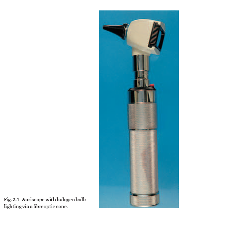
The ear is most conveniently examined with an auriscope (Fig. 2.1).
Modern auriscopes have distal illumination via a fibre-optic cone giving a
bright, even light. Because interpretation of the appearance depends to a
5
Fig. 2.1 Auriscope with halogen bulb
lighting via a fibreoptic cone.
Download Files
Course Material
- The Ear: Some Applied Anatomy
- Clinical Examination of the Ear
- Testing the Hearing
- Deafness
- Conditions of the Pinna
- Conditions of the External Auditory Meatus
- Injury of the Tympanic Membrane
- Acute Otitis Media
- Chronic Otitis Media
- Complications of Middle-Ear Infection
- Otitis Media with Effusion
- Otosclerosis
- Earache (Otalgia)
- Vertigo
- Facial Nerve Paralysis
- Adenoids
- The Tonsils and Oropharynx
- Tonsillectomy
- Retropharyngeal Abscess
- Examination of the Larynx
- Injuries of the Larynx and Trachea
- Acute Disorders of the Larynx
- Chronic Disorders of the Larynx
- Tumours of the Larynx
- Vocal Cord Paralysis
- Airway Obstruction in Infants and Children
- Conditions of the Hypopharynx
- Tracheostomy
- Diseases of the Salivary Glands
- Chapters 29
- Department Sargodha Medical College
- Teacher
Dr. Muhammad Khalil


