
The Ear: Some Applied Anatomy
THE PINNA
The external ear or pinna, is composed of cartilage with closely adherent
perichondrium and skin. It is developed from six tubercles of the first
branchial arch. Fistulae and accessory auricles result from failure of fusion
of these tubercles.
THE EXTERNAL AUDITORY MEATUS
The external auditory meatus is about 25 mm in length, has a skeleton
of cartilage in its outer third (where it contains hairs and ceruminous
glands) and has bone in its inner two-thirds. The skin of the inner part is
exceedingly thin, adherent and sensitive. At the medial end of the meatus
there is the antero-inferior recess, in which wax, debris or foreign bodies
may lodge.
THE TYMPANIC MEMBRANE (Fig. 1.1)
The tympanic membrane is composed of three layer—skin, fibrous tissue
and mucosa.The normal appearance of the membrane is pearly and opaque,
with a well-defined light reflex due to its concave shape.
THE TYMPANIC CAVITY
Medial to the tympanic membrane, the tympanic cavity is an air-containing
space 15 mm high and 15 mm antero-posteriorly, although only 2mm deep
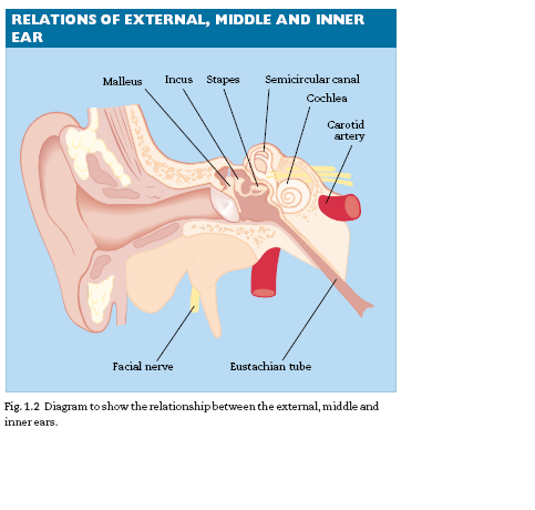
in parts. The middle ear contains the ossicular chain of malleus, incus and
stapes (Fig. 1.2) and its medial wall is crowded with structures closely related
to one another: the facial nerve, the round and oval windows, the lateral
semicircular canal and basal turn of the cochlea.The major reason for having
an air-containing middle ear is to reduce the acoustic impedance that
would be caused if a sound wave in air were to be applied directly to the
cochlear fluids.Without this impedance matching, 99% of the sound energy
would simply be reflected at an air/fluid interface.
THE EUSTACHIAN TUBE
The Eustachian tube connects the middle-ear cleft with the nasopharynx
and is responsible for the aeration of the middle ear. The tube is morehorizontal in the infant than in the adult and secretions or vomit may enter
the tympanic cavity more easily in the supine position. The tube is normally
closed and is opened by the palatal muscles on swallowing.This is impaired
by the presence of a palatal cleft.
THE FACIAL NERVE
The facial nerve is embedded in bone in its petrous part but exits at the stylomastoid
foramen (Fig. 1.3). In infants, the mastoid process is undevelopedand the nerve very superficial.


Download Files
Course Material
- The Ear: Some Applied Anatomy
- Clinical Examination of the Ear
- Testing the Hearing
- Deafness
- Conditions of the Pinna
- Conditions of the External Auditory Meatus
- Injury of the Tympanic Membrane
- Acute Otitis Media
- Chronic Otitis Media
- Complications of Middle-Ear Infection
- Otitis Media with Effusion
- Otosclerosis
- Earache (Otalgia)
- Vertigo
- Facial Nerve Paralysis
- Adenoids
- The Tonsils and Oropharynx
- Tonsillectomy
- Retropharyngeal Abscess
- Examination of the Larynx
- Injuries of the Larynx and Trachea
- Acute Disorders of the Larynx
- Chronic Disorders of the Larynx
- Tumours of the Larynx
- Vocal Cord Paralysis
- Airway Obstruction in Infants and Children
- Conditions of the Hypopharynx
- Tracheostomy
- Diseases of the Salivary Glands
- Chapters 29
- Department Sargodha Medical College
- Teacher
Dr. Muhammad Khalil
