
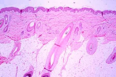
Histology of Skin
Skin is part of the integumentary system and considered to be the largest organ of the human body. There are two layers of skin: the epidermis, the dermis, and the hypodermis (subcutaneous fat) not a part of skin.
The first and outermost layer of skin is the epidermis. The epidermis is a stratified squamous keratinized epithelium that contains four to five layers depending on its location.
There are four types of cells.
- - Keratinoytes
- - Melanocytes
- - Merkel cells
- - Langerhans’ cells
Deep to the epidermis lies the dermis. It is a thick layer of dense connective tissue consisting of collagen and elastic fibers. The dermis also contains nerve endings, blood vessels, hair shafts, sweat glands, and sebaceous glands. The apical layer of dermis folds to form papillae that extend into the epidermis like tiny finger-like projections and is referred to as the papillary dermis, while the lower layer of the dermis is referred to as the reticular dermis.
The hypodermis is the third and deepest layer consisting mainly of adipose tissue.
Objective:
- Identify principal layers of the skin (epidermis, dermis and hypodermis) at the light microscope
- Identify the layers of the epidermis in thick and thin skin.
- Identify melanocytes and keratinocytes.


Download Files
- Chapters 9
- Department Anatomy
- Teacher
Dr. Muhammad Aaqib Riaz
