
WEEK 4:NEUROMUSCULAR JUNCTION
Transmission of Impulses
from Nerve Endings to
Skeletal Muscle Fibers:
The Neuromuscular
Junction:
The skeletal muscle fibers are innervated by large,
myelinated nerve fibers that originate from large motoneurons in the anterior
horns of the spinal cord. Each nerve fiber, after
entering the muscle belly, normally branches and stimulates from three to
several hundred skeletal muscle fibers. Each nerve ending makes a junction,
called the neuromuscular junction, with the muscle fiber near its midpoint. The
action potential initiated in the muscle fiber by the nerve signal travels in both
directions toward the muscle fiber ends.With the exception of about 2 per cent
of the muscle fibers, there is only one such junction per muscle fiber.
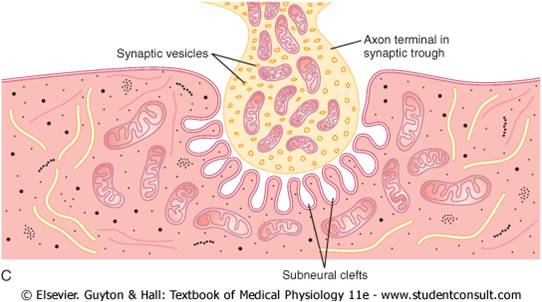
Physiologic Anatomy of the Neuromuscular Junction—The Motor End Plate-The nerve fiber forms a complex of branching nerve
terminals that invaginate into the surface of the muscle fiber but lie outside the
muscle fiber plasma membrane. The entire structure is called the motor end
plate. It is covered by one or more Schwann cells that insulate it from the
surrounding fluids. The invaginated membrane
is called the synaptic gutter or synaptic trough, and the space between the
terminal and the fiber membrane is called the synaptic space or synaptic cleft.
This space is 20 to 30 nanometers wide. At the bottom of the gutter are numerous
smaller folds of the muscle membrane called subneural clefts, which greatly
increase the surface area at which the synaptic transmitter can act.
In the axon terminal are many mitochondria that supply adenosine triphosphate
(ATP), the energy source that is used for synthesis of an excitatory
transmitter acetylcholine. The acetylcholine in turn excites the muscle
fiber membrane. Acetylcholine is synthesized in the cytoplasm of the terminal,
but it is absorbed rapidly into many small synaptic vesicles, about 300,000
of which are normally in the terminals of a single end plate. In the synaptic
space are large quantities of the enzyme acetylcholinesterase, which destroys
acetylcholine a few milliseconds after it has been released from the synaptic vesicles.
The formation
and release of acetylcholine at this junction occur in the
following stages:
1. Small vesicles, about 40 nanometers in size, are
formed by the Golgi apparatus in the cell body of
the motoneuron in the spinal cord. These vesicles
are then transported by axoplasm that “streams”
through the core of the axon from the central
cell body in the spinal cord all the way to the
neuromuscular junction at the tips of the peripheral
nerve fibers. About 300,000 of these small vesicles
collect in the nerve terminals of a single skeletal
muscle end plate.
2. Acetylcholine is synthesized in the cytosol of the
nerve fiber terminal but is immediately transported
through the membranes of the vesicles to their
interior, where it is stored in highly concentrated
form, about 10,000 molecules of acetylcholine in
each vesicle.
3. When an action potential arrives at the nerve
terminal, it opens many calcium channels in the
membrane of the nerve terminal because this
terminal has an abundance of voltage-gated
calcium channels. As a result, the calcium ion
concentration inside the terminal membrane
increases about 100-fold, which in turn increases
the rate of fusion of the acetylcholine vesicles with
the terminal membrane about 10,000-fold. This
fusion makes many of the vesicles rupture, allowing
exocytosis of acetylcholine into the synaptic space.
About 125 vesicles usually rupture with each
action potential. Then, after a few milliseconds, the
acetylcholine is split by acetylcholinesterase
into acetate ion and choline, and the choline is
reabsorbed actively into the neural terminal to be
reused to form new acetylcholine. This sequence of
events occurs within a period of 5 to 10
milliseconds.
4. The number of vesicles available in the nerve
ending is sufficient to allow transmission of only a
few thousand nerve-to-muscle impulses. Therefore,
for continued function of the neuromuscular
junction, new vesicles need to be re-formed rapidly.Within a few seconds after each action potential is
over, “coated pits” appear in the terminal nerve
membrane, caused by contractile proteins in the
nerve ending, especially the protein clathrin, which
is attached to the membrane in the areas of the
original vesicles.Within about 20 seconds, the
proteins contract and cause the pits to break away
to the interior of the membrane, thus forming new
vesicles.Within another few seconds, acetylcholine
is transported to the interior of these vesicles, and
they are then ready for a new cycle of acetylcholine
release.


Download Files
- Chapters 5
- Department Physiology
- Teacher
Dr. Madiha Nazir
