
WEEK 2:SKELETOL MUSCLE
The initiation and execution of muscle contraction
occur in the following sequential steps:
1. An action potential travels along a motor nerve to
its endings on muscle fibers.
2. At each ending, the nerve secretes a small amount
of the neurotransmitter substance acetylcholine.
3. The acetylcholine acts on a local area of the muscle
fiber membrane to open multiple “acetylcholinegated”
channels through protein molecules floating
in the membrane
.4. Opening of the acetylcholine-gated channels allows
large quantities of sodium ions to diffuse to the
interior of the muscle fiber membrane. This initiates
an action potential at the membrane.
5. The action potential travels along the muscle fiber
membrane in the same way that action potentials
travel along nerve fiber membranes.
6. The action potential depolarizes the muscle
membrane, and much of the action potential
electricity flows through the center of the muscle
fiber. Here it causes the sarcoplasmic reticulum to
release large quantities of calcium ions that have
been stored within this reticulum.
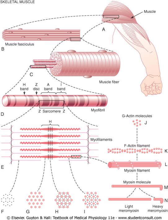
7. The calcium ions initiate attractive forces between
the actin and myosin filaments, causing them to
slide alongside each other, which is the contractile
process.
8. After a fraction of a second, the calcium ions are
pumped back into the sarcoplasmic reticulum by a
Ca++ membrane pump, and they remain stored in
the reticulum until a new muscle action potential
comes along; this removal of calcium ions from the
myofibrils causes the muscle contraction to cease.
Muscle Hypertrophy and Muscle Atrophy- When the total
mass of a muscle increases, this is called muscle hypertrophy.
When it decreases, the process is called muscle
atrophy.
Virtually all muscle hypertrophy results from an
increase in the number of actin and myosin filaments in
each muscle fiber, causing enlargement of the individual
muscle fibers; this is called simply fiber hypertrophy.
Hypertrophy occurs to a much greater extent when the
muscle is loaded during the contractile process. Only a
few strong contractions each day are required to cause
significant hypertrophy within 6 to 10 weeks.
The manner in which forceful contraction leads to
hypertrophy is not known. It is known, however, that the
rate of synthesis of muscle contractile proteins is far
greater when hypertrophy is developing, leading also to
progressively greater numbers of both actin and myosin
filaments in the myofibrils, often increasing as much as
50 per cent. In turn, some of the myofibrils themselves
have been observed to split within hypertrophying
muscle to form new myofibrils, but how important this
is in usual muscle hypertrophy is still unknown.
Along with the increasing size of myofibrils, the
enzyme systems that provide energy also increase. This
is especially true of the enzymes for glycolysis, allowing
rapid supply of energy during short-term forceful
muscle contraction.
When a muscle remains unused for many weeks, the
rate of decay of the contractile proteins is more rapid
than the rate of replacement.Therefore, muscle atrophy
occurs.
Hyperplasia of Muscle Fibers- Under rare conditions of
extreme muscle force generation, the actual number of
muscle fibers has been observed to increase (but only
by a few percentage points), in addition to the fiber
hypertrophy process. This increase in fiber number is
called fiber hyperplasia. When it does occur, the mechanism
is linear splitting of previously enlarged fibers.
Rigor Mortis:
Several hours after death, all the muscles of the body go
into a state of contracture called “rigor mortis”; that is,
the muscles contract and become rigid, even without
action potentials.This rigidity results from loss of all the
ATP, which is required to cause separation of the crossbridges
from the actin filaments during the relaxation
process. The muscles remain in rigor until the muscle
proteins deteriorate about 15 to 25 hours later, which
presumably results from autolysis caused by enzymes
released from lysosomes. All these events occur more
rapidly at higher temperatures.


Download Files
- Chapters 5
- Department Physiology
- Teacher
Dr. Madiha Nazir
