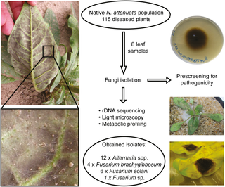
Practical Isolation of Plant pathogenic Fungi
ISOLATION OF FUNGAL PATHOGENS
3a.Isolation of plant pathogenic fungi from diseased plant material:
Isolation of the fungal pathogens from diseased material is made by surface
sterilizing the diseased area with surface sterilizing agents, removing a small portion of
the infected tissue (leaves, stems, fruits etc.) with a sterile scalpel, and plating it in a
plate containing a nutrient medium. The most common method, for isolating fungal
pathogens from infected leaves as well as other plant parts involves cutting several small
sections 5-10 mm-square from the margin of the infected lesion to contain both diseased
and healthy looking tissue. These are placed in surface sterilizing agents solutions for
about 15-30 seconds the sections are taken out aseptically and blotted dry on clean,
sterile paper towels or washed in three changes of sterile water and are finally placed on
the nutrient medium, usually three to five per dish. The pathogen will grow from the
sections and the colonies of the pathogen are sub cultured aseptically for further study.
Materials required
Infected young leaves, , sterile Petri -dishes, PDA slants, sodium hypochlorite
solution ( 1 % ), sterile water, razor blade, forceps, inoculation needle, burner/spirit lamp,
spirit, incubator, PDA medium.
Procedure
1. Select infected host tissue from the advancing margin of the lesions.
2. Cut into small pieces (2-5 mm ) containing both the diseased and healthy tissue
and keep in sterile Petri dishes
3. Dip the pieces into 1 % sodium hypochlorite solution for about one minute.
4. Transfer the pieces to Petri - dishes containing sterile distilled water and wash
thoroughly in two changes of sterile water to free them from the chemicals if any.
5. Wash hands with rectified spirit and wipe the table top of inoculation chamber-'
with rectified spirit.
6. Lit the burner
7. Hold the flask containing sterile Luke warm PDA in the right hand and remove
plug near the flame. Lift the lid of Petri dish gently with left hand and pour about
20 ml of medium. Close the mouth of the flask with plug near the flame
8. After solidification of the medium, place four sterilized pieces at different
distance in a single PDA plate.
9. Incubate the Petri dishes in an inverted position at 25° C and examine for 3-5
days.
Observations and results
Observe the incubated plates from the second day onwards for the growth of the
fungus. Aseptically transfer the bits of mycelia from the margin of the colonies on fresh
PDA slants for further study. Mycelia growth on the medium from the infected tissues,
indicates that the disease may be due to a fungus.


