
Practical Isolation of Plant pathogenic Bacteria
Isolation of phyto-pathogenic bacteria from diseased plants
Isolation and identification of bacteria associated with diseased plant is important
to determine whether bacteria are involved in plant disease. The method normally used
to isolate phyto-pathogenic bacteria differs from that used for fungi. A suspension of
bacterium is prepared from the infected material and loopfuls of this are streaked onto
nutrient agar plates. The aim is to produce single colony that can be sub-cultured pure.
Pure cultures are absolutely essential for pathogenicity assays and characterizing the
pathogen for identification. The serial dilution method is used for isolating bacteria from
diseased tissues contaminated with other bacteria. After surface sterilization of sections
of diseased tissues, the sections are ground in small volumes of sterile water and then
part of this homogenate is diluted serially. Finally, plates containing nutrient agar are
streaked with a loop dipped in each of the different serial dilutions and single colonies of
the pathogenic bacterium are obtained from the higher dilutions that still contain bacteria.
Choice of material: Selection of the diseased tissue is important because pathogenic
bacteria may occupy different locations in the plant. In isolation of bacteria, it is generally
better to use newly collected material. The earliest stages of symptom development
should be used. Old lesions and dead areas usually contain few pathogens and many
saprophytes. Necrotic diseases usually start with tiny, dark greenish, spots, which are
excellent for isolations. Cankers and soft rots should either be at an early stage, or if,
older lesions only are available, the advancing edge must be used, where the disease is
spreading into healthy tissue. When crown gall is suspected in a woody plant a search
must be made for young galls on young green stems. With wilts and other vascular
infections small pieces of infected stem are usually good for isolation.
Preparation of material: Clean leaves and stems, carefully chosen and "handled
aseptically, can often be used without surface sterilization. Roots and parts contaminated
with soil should be gently washed with clean water as soon as possible after collection.
Medium: Nutrient agar is suitable for the isolation of most plant pathogens. The medium
used for isolations must have a dry surface. If water is present the bacteria move around
and a carpet of mixed growth results instead of the required single colony.
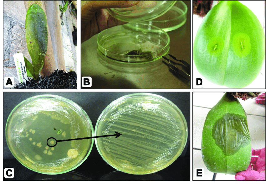
This exercise deals with the isolation of bacterium, Xanthomonas axonopodis pv.
citri causal agent of citrus canker.
Materials
Fresh citrus leaves infected by Xanthomonas axonopodis pv. citri, nutrient agar
medium, surface sterilizing agents (1 % sodium hypochlorite), sterile razor blade, glass
rod, sterile water, sterile test tubes and Petri-dishes, sterile pipettes (I ml), inoculation
loop.
Procedure
Put on the U.V lamp of inoculation chamber for 5 mts. Wipe the table top with
rectified spirit Wash hands with rectified spirit and air dry. Lit the burner or spirit lamp,
arrange sterile Petri dishes near the burner.
1. Select a diseased citrus leaf infected by canker and cut out a small portion of the
diseased tissue from the advancing lesion using sterile razor blade in a drop of
sterile water and after several minutes, examine under microscope . If bacterial
ooze is seen, proceed for isolation
Surface disinfests the cut portions by dipping in sodium hypochlorite solution for 60
sec. and then immediately rinse three times with sterile water.
3. Immerse the disinfested cut portions in I ml of sterile water taken in a clean
sterilised test tube.
4. Crush the cut portions of the leaf with a sterile glass rod. Allow it to stand for 5
minutes to allow the bacteria to diffuse out of the cut tissue and into the water.
5. Gently lift the lid of a Petri dish with left hand and using inoculation loop transfer
several loopfuls of the bacterial suspension to sterile Petri-dishes (three) containing
1 ml of sterile water and mix thoroughly.
6. Hold flask filled with sterile Luke warm nutrient agar medium in the right hand and
remove cotton plug near the flame and pour about 20 ml of medium into each dish
and mix thoroughly by gentle rotation. Allow time for solidification of medium.
7. Incubate the dishes in an inverted position at 25°C for 36 to 72 hours.
8. Observation: Observe the dishes for appearance of desired bacterial colonies. If
colonies appear, select consistently found and well isolated colonies of the
pathogen, for sub-culturing and further studies.
9. Select the isolated colonies and streak on the surface of a solidified medium in a
zigzag manner and incubate the dishes at 25oC. Bacteria isolated from nature may
be contaminated with saprophytic species; hence, re-streaking for isolation ensures
a pure culture. Transfer some of the purified colonies to NA slants and grow them
for further use.
EXERCISE
Follow the protocol outlined in the above procedure and isolate the desired bacterial
colonies from the given diseased material and maintain pure culture in Petri dishes and
slants for further study.


Download Files
- Leaf or Brown Rust of Wheat
- Stripe or Yellow Rust of Wheat
- Rice Blast
- Foot Rot (Bakanae Disease of Rice)
- Loose Smut of Wheat
- Flag Smut of Wheat
- Old Bunt of Wheat
- New Bunt of Wheat (Karnal Bunt of Wheat)
- Ear Cockle of Wheat
- Powdery Mildew of Wheat
- Take All of Wheat
- Rice Hoja Blanca
- Fusarium Wilt of Cotton
- Root Rot of Cotton
- Charcol Rot of Maize
- Boll Rot of Cotton
- Practical Demonstration of Koch's Postulates for Fungi
- Practical Demonstration of Koch's Postulates for Bacteria
- Practical Preparation of Herbarium
- Practical Diagnosis of Wheat and Barley Diseases
- Practical Types of Media
- Practical Preparation of Media
- Practical Isolation of Plant pathogenic Fungi
- Practical Isolation of Plant pathogenic Bacteria
- Practical Plant Disease Diagnostics
- Practical Moist Chamber Incubation
- Chapters 26
- Department Plant Pathology
- Teacher
Dr. Muhammad Ahmad Zeshan
