
Week6 The Viruses: Introduction and cultivation
Viruses Introduction and Cultivation
These are obligate intracellular parasites. That is, they absolutely require living host cells in order to multiply. Because viruses are inert outside living host cells, in this sense they are not considered to be living organisms. However, once viruses enter a host cell, the viral nucleic acids become active, and viral multiplication results. In this sense, viruses are alive when they multiply in the host cells they infect. From a clinical point of view, viruses can be considered alive because they cause infection and disease.
Viruses have few or no enzymes of their own for metabolism; for example, they lack enzymes for protein synthesis and ATP generation. To multiply, viruses must take over the metabolic machinery of the host cell. This fact has considerable medical significance for the development of antiviral drugs, because most drugs that would interfere with viral multiplication would also interfere with the functioning of the host cell and therefore are too toxic for clinical use. Viruses are completely unaffected by antibiotics because they lack a cell wall, ribosomes and other structures.
Many researchers are trying to develop more effective antiviral drugs
The drugs available are only effective against a specific type of virus. They prevents the virus from entering host cell, some drugs interfere with uncoating step so prevents replication
examples: Amantadine and Rimantadine block the uncoating of influenza A virus after it enters the cell. To prevent production and release of viral particles are the targets of some antiviral medications.
The viral infected cell can prepare neighboring cells against a potential infection by the virus by releasing interferon. In response to interferon, cells produce large amounts of antiviral proteins which include oligoadenylate synthetase and protein kinase. Interferon action destroys RNA within the cells to further reduce protein synthesis of both viral and host genes. Inhibited protein synthesis destroys both the virus and infected host cells. In addition, interferons induce production of hundreds of other proteins known collectively as interferon-stimulated genes (ISGs) that have roles in combating viruses and other actions produced by interferon.
Size: 10-100 times smaller than most bacteria, with approximate size of 20-1000nm.
Host Range: Vertebrates, invertebrates, plants, fungi, bacteria and protists.
Structure: A viron is complete, fully-developed infectious particle, composed of Nucleic Acid surrounded by protein that protects it from environment. A virus can has either DNA or RNA but never both. A nucleic acid can be single or double stranded which can be either linear or circular. Total amount of Nucleic Acid varies from 2,000 nucleotides to 2, 50,000 Nucleotides. A nucleic acid of virus is surrounded by protein coat called capsid, which is composed of protein subunits called capsomere. In some virus capsid is covered by envelop which consist of combination of lipids, proteins and carbohydrates. Depending upon virus envelop may or may not be covered by spikes. Spikes are protein carbohydrate complex that projects from surface of envelop and these are means of attachment of virus to host cell. Tail is also present in some viruses while it is absent in others. It also helps viral attachment to different surfaces.
Morphology Virus may be classified into several different morphological types on bases of their capsid architect.
1.Helical or Rod Shaped:
Some viruses reassemble long rod that may be rigid or flexible e.g. Rabies virus.
2. Polyhedral Virus
Many animal/plant and bacterial viruses are polyhedral or many sided virus e.g. Polio and Adenovirus
3. Spherical
Enveloped viruses are roughly spherical e.g. influenza virus.
4. Complex Viruses
Some virus e.g. bacteriophage have complicated structure and are called complex viruses. These viruses have polyhedral head and helical tail e.g. T. even bacteriophage virus.
Viral Cultivation
- Bacterial Viruses :
Viruses are easily isolated cultivated in young actively growing culture of bacteria in broth or
on agar plate.
- Broth Culture
In broth culture the lysying bacteria may cause a cloudy culture to become clear.
- Agar Plates
A sample of bacteriophage is mixed with host bacteria on agar media which are then poured in Petri plates containing hardened layer of agar-broth medium. A viral-bacterial mixture is solidified in thin top layer. Each Virus impacts a bacterium multiplies and release several new viruses that infects other bacteria so all bacteria in area are destroyed which produce number of clearing or plaque visible against a lawn of bacterial growth on surface of agar. Each plaque corresponds to a single virus so concentration of viral suspension is measured by number of plaques in term of plaque forming units.
Plant Viruses:
Plant viruses can be cultivated by direct inoculation of viral suspension by rubbing on leaves of living plants accompanied by abrasion which leads to formation of local lesions and general infections. Cell culture has also been developed for cultivating the plant virus. e.g. Rhabdovirus has been developed on leaves hopper cell culture.
Growing Animal Viruses in the laboratory
In the laboratory, three methods are commonly used for culturing animal viruses. These methods involve using living animals, embryonated eggs, or cell cultures.
In living Animals
Some animal viruses can be cultured only in living animals, such as mice, rabbits, and guinea pigs. Most experiments to study the immune system's response to viral infections must also be performed in virally infected live animals. Animal inoculation may be used as a diagnostic procedure for identifying and isolating a virus from a clinical specimen. After the animal is inoculated with the specimen, the animal is observed for signs of disease or is killed so that infected tissues can be examined for the virus.
Some human viruses cannot be grown in animals or if can be grown do not cause disease.
In Embryonated Eggs
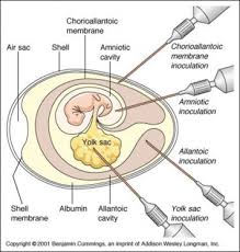
If the virus will grow in an embryonated egg, this can be a fairly convenient and inexpensive form of host for many animal viruses. A hole is drilled in the shell of the embryonated egg, and a viral suspension or suspected virus-containing tissue is injected into the fluid of the egg. There are several membranes in an egg, and the virus is injected near the one most appropriate for its growth. Viral growth is signaled by the death of the embryo by embryo cell damage, or by the formation of typical pocks or lesions on the egg membranes. This method was once the most widely used method of viral isolation and growth, and it is still used to grow viruses for some vaccines. For this reason, you may be asked if you are allergic to eggs before receiving a vaccination, because egg proteins may be present in the viral vaccine preparations.
In Cell Cultures
Cell cultures have replaced embryonated eggs as the preferred type of growth medium for many viruses. Cell cultures consist of cells grown in culture media in the laboratory. Because these cultures are generally rather homogeneous collections of cells and can be propagated and handled much like bacterial cultures, they are more convenient to work with than whole animals or embryonated eggs.
Cell culture lines are started by treating a slice of animal tissue with enzymes that separate the individual cells. These cells are suspended in a solution that provides the osmotic pressure, nutrients, and growth factors needed for the cells to grow. Normal cells tend to adhere to the glass or plastic container and reproduce to form a monolayer.
Viruses may be grown in primary or continuous cell lines. Primary cell lines, derived from tissue slices, tend to die out after only a few generations. Certain cell lines, called diploid cell lines, developed from human embryos can be maintained for about 100 generations and are widely used for culturing viruses that require a human host. Cell1ines developed from embryonic human cells are used to culture rabies virus for a rabies vaccine called human diploid culture vaccine.
When viruses are routinely grown in a laboratory, continuous cell lines are used. These are transformed (cancerous) cells that can be maintained through an indefinite number of generations, and they are sometimes called immortal cell lines. One of these, the HeLa cell
line, was isolated from the cancer of a woman (Henrietta Lacks) who died in 1951. After years of laboratory cultivation, many such cell lines have lost almost all the original characteristics of the cell. There are still some viruses that have never been successfully cultivated in cell culture. A major problem with cell culture is that the cell lines must be kept free of microbial contamination. The maintenance of cell culture lines requires trained technicians with considerable experience working on a full -time basis. Because of these difficulties, most hospital laboratories and many state health laboratories do not isolate and identify viruses in clinical work


Download Files
- Week I General Microbiology
- Week 2 The Morphology and Fine Structure of Bacteria
- Week 3 Classification of Bacteria
- Week 4 Bacterial Growth and Nutrition
- Week 5 Bacterial culture Media and staining methods
- Week6 The Viruses: Introduction and cultivation
- The Viruses Classification and multiplication
- Mid term Examination
- Cytopathic effects of viruses/ Oncogenic viruses
- Fungi
- Protozoa Morphology, Importance, Classification
- Protozoal diseases
- Normal Flora
- Microbiology of Air
- Aquatic Microbiology
- Final term Examination
- Chapters 16
- Department College of Pharmacy
- Teacher
Dr. Alia Erum
